Unoffiical Guide to Radiology 100 CXR

The Unofficial Guide to

Radiology:

100 Practice Chest X-Rays,with Full Colour Annotations and Full X-Ray Reports

Associate Editors: Mohammed Rashid Akhtar, Na’eem Ahmed and Nihad Khan

Series Editors: Mark Rodrigues & Zeshan Qureshi

Buy Now

“This is probably the easiest way of learning the basics of chest x-ray interpretation. An excellent introduction for the beginner and a superb way of revising the subject for those of us who are rather rusty.”

Dr David Wilson
President of the British Institute of Radiology

“Each image is presented clearly with the relevant anatomical features and abnormalities highlighted clearly and set in the context of the pathophysiology. I’m sure that those who read this book will never be left standing in silence at the dreaded radiology OSCE station!”

Simon Maxwell testimonial image

Simon Maxwell
Professor of Student Learning, University of Edinburgh

“The clarity of the on-image labelling gives immediate feedback, enabling the reader to make sense of each radiograph. I wish I’d had a copy when I was a medical student.”

Bob Clarke photo

Bob Clarke
Associate Dean, Professional Development, London.
Director, Ask Doctor Clarke Ltd.

“The book will provide doctors with more confidence and competence and, more importantly, it addresses a patient safety issue in that missing something on an X-Ray could have important implications for patients and their well-being. Congratulations on another excellent book in the series which we’ll be certain to let our Graduate Entry Medical Students at Swansea know about.”

Prof Judy McKimm

Professor Judy McKimm
Professor of Medical Education and Director of Strategic Educational Development, University of Swansea

“The wealth of high quality teaching material in this carefully curated collection of 100 chest radiographs is impressive, and the authors have rightly concentrated on common, but clinically important, conditions.”

David M Hansell
Professor of Thoracic Imaging, Imperial College, London

“I only wish I had access to this book in my first few years on clinical placement. The high quality images, clearly labelled pathological signs and broad range of chest pathology covered, make this book an invaluable tool to anyone looking to develop a solid foundation in interpreting chest x-rays.”

Lana Nguyn

Lana Nguyen
President, Western Sydney Medical Society 2015-6

“The book is fantastic. It is so concise and reinforces learning through repetition and clear descriptions. A perfect compendium for the clinical medical student.”

William Rees Photo

William Ries
President, Students Section, Royal Society Medicine

“This book helps to make the process of interpreting X-rays easy to learn and memorable. There are lots of useful tips and several examples which vary in complexity so you can test yourself as you go along. I would recommend this to any medical student.”

Dvolka Ganesh

Divolka Ganesh
BSc in Medical Imaging, Year 4 Medical Student, University of Leeds

“Logical, has clear images and leads you to consider all the elements of interpreting radiographs. Case-based scenarios means it has clinical relevance that is really helpful for learning how to incorporate imaging into managing common pathologies.”

Catherine Lovegrove photo

Catherine Elizabeth Lovegrove
BSc (Hons.), Final Year Medical Student, King’s College London

“Fantastic resource-much needed. This guide allows students to test themselves, spot pointers and then have a full explanation of the pathology and radiological findings. I also loved the difficulty ratings from easy to hard, this is a great way to learn and build on your knowledge base.”

Lauren Quinn
Medical Student, University of Birmingham

“Even when you’re done with medical school it’s really important to carry on assessing X-rays, especially for specialities you might not be working in.. The information in the books is accessible, clearly laid out and is great for new concepts as well as reinforcing the stuff you already know.”

Harriet Bedell Pearce photo

Harriet Bedell-Pearce
Junior Doctor

“An excellent workbook for medical students which is clear and concise. All the radiographs are large and well pictured and the explanations are written out colour-coded which is an excellent way to learn and memorise the details. Would be invaluable to help with interpretations of x-rays in OSCEs.”

Arwa Alanizi
Final year medical student at KCL

“The x-ray cases are clearly annotated and colour coded and you are led through each case in a clear systematic way. This repetition allows you to build up confidence for presenting a range of scenarios to colleagues. I thoroughly recommend this book.”

Isabelle Guest
Fourth year medical student, St George’s University

“A fantastic resource! The layout is perfect for testing yourself, and the feedback is systematic, clear and concise. The use of colour to highlight points on the chest x-ray is particularly useful. There are a variety of cases, which become increasingly complex, so a great book for any stage in medical school.”

Alice Southwell photo

Alice Southwell
Fourth Year Medical Student, University of Leeds

“The similarities between this and the Unofficial Guide to Radiology book are the case descriptions with imaging on one page, followed by an entire page of markings that we should have spotted on the next page. This technique allows us to think for ourselves rather than cheating our way through, like most other textbooks do.”

Pooja Parekh

Pooja Parekh
Junior Doctor, Great Ormond Street Hospital, UK

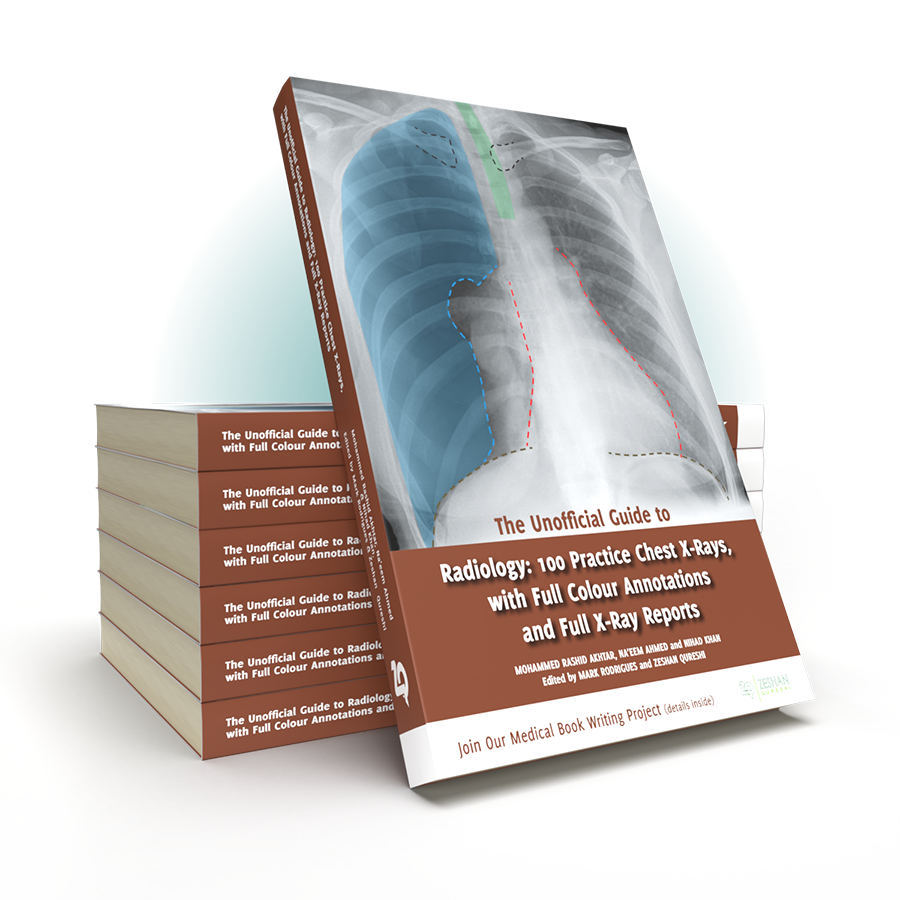
Following on from the multi-award winning Unofficial Guide to Radiology, the Unofficial Guide team return with a practical work book on chest X-rays interpretation, giving you all the practice you need to master this vital skill.

Despite its universal importance, X-ray interpretation is often an overlooked subject in the medical school curriculum, making it difficult and daunting for many medical students and junior doctors.

The Unofficial Guide to Radiology: 100 Practice Chest X-Rays, with Full Colour Annotations and Full X-Ray Reports aims to help address this. This follow-up textbook, specifically designed for medical students, radiographers, physician’s associates, and junior doctors, builds upon these foundations, providing readers with the opportunity to practise and consolidate their chest X-ray assessment and presenting skills.

Key Features

Written in line with the other books in the series, stations contain:

100 large, high quality chest X-rays to assess.
j
Cases presented in the context of a clinical scenario and covering a wide range of common and important findings (in line with the Royal College of Radiologists’ Undergraduate Radiology Curriculum)
Detailed on-image colour annotations to highlight key findings
Comprehensive systematic X-ray reports
Relevant further investigations and management are discussed for each case
The Unofficial Guide to Radiology: 100 Chest X-rays is an invaluable resource for anyone hoping to gain a practical understanding and a wealth of experience of chest X-rays and their interpretation in a concise, student-focused manner.
Zeshan Qureshi
Zeshan Qureshi
Chief Editor
Paediatrics trainee at Great Ormond Street, and the Institute of Child Health

With this textbook, we hope you will become more confident and competent interpreting chest X-rays, both in exam situations and in clinical practice.

We want you to get involved! This textbook has been a collaboration with junior doctors and students just like you. You have the power to contribute something really valuable to medical education; we welcome your suggestions and would love for you to get in touch.

A good starting point is “The Unofficial Guide to Medicine” facebook page, an active forum for medical education. Please get in touch and be part of the medical education project.

100 Chest X-rays: Reported and Annotated JUST LIKE THIS

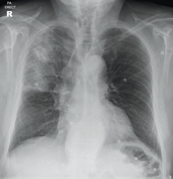
A 70 year old male who lives in a residential home presents to ED with increasing confusion. He has a productive cough and a fever. He has a past medical history of hypertension, angina and mild cognitive impairment. He has a 25 pack year smoking history. On examination, he has saturations of 89% in air, and is febrile with a temperature of 38.8°C. There is dullness to percussion and coarse crackles in the right upper zone. A chest X-ray is requested to assess for possible pneumonia or collapse.

Can you report the X-ray and put in place a management plan?

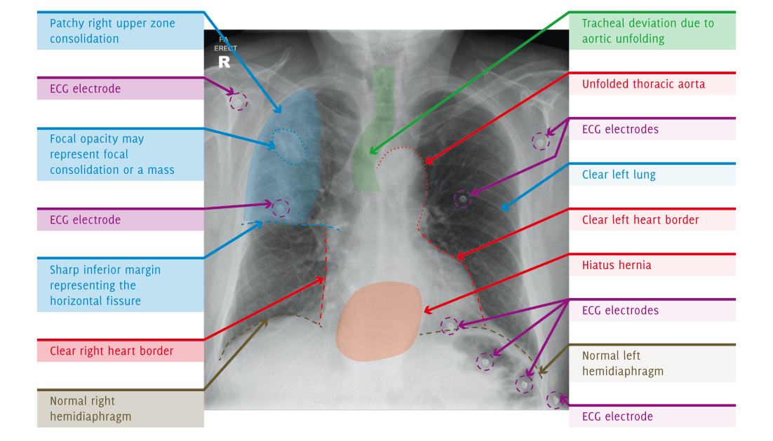
Turning over the page reveals a full on-image colour annotated X-ray, along with the report, detailing everything you should be expected to see on the X-ray.

The Unoffical Guide To Radiology - X-Ray

Full Report Covering Every Detail Of The X-Ray You Need To Know

Patient ID: Anonymous
Projection: PA
Penetration: Adequate – vertebral bodies just visible behind heart
Inspiration: Adequate – 8 anterior ribs visible
Rotation: Not rotated

AIRWAY

The upper trachea is central. The lower trachea is displaced to the right by the aortic arch.

BREATHING

There is heterogeneous air space opacification in the right upper zone. This has a relatively well defined inferior margin, which is likely to represent the horizontal fissure. There is a focal area of increased opacification in the right upper zone, which may represent focal consolidation or an underlying mass. The remainder of the lungs are clear. The lungs are not hyperinflated.
The pleural spaces are clear.
Normal pulmonary vascularity.

CIRCULATION

The heart is not enlarged.
The heart borders are clear.
There is unfolding of the thoracic aorta, which displaces the lower trachea to the right.
The mediastinum is central, not widened, with clear borders. There is a welldefined density projected over the lower mediastinum, which is in keeping with a hiatus hernia.
Normal size, shape, and position of both hila.

DIAPHRAGM + DELICATES

Normal appearance and position of hemidiaphragms.
No pneumoperitoneum.
The imaged skeleton is intact with no fractures or destructive bony lesions visible.
The visible soft tissues are unremarkable.

EXTRAS + REVIEW AREAS

ECG electrodes in situ.
No vascular lines, tubes or surgical clips.
Lung Apices: Heterogeneous right apical consolidation. Normal left apex
Hila: Normal
Behind Heart: There is a retrocardiac density, which represents a hiatus hernia
Costophrenic Angles: Normal
Below the Diaphragm: Normal

Fully Annotated X-Ray, With On Image, Colour Labeling For Unbeatable Clarity

Annotated X-Ray image

SUMMARY, INVESTIGATIONS & MANAGEMENT

This X-ray demonstrates heterogeneous right upper zone consolidation in keeping with pneumonia. The consolidation has a relatively abrupt inferior margin in keeping with the horizontal fissure, indicating this is right upper lobe pneumonia. A focal opacity in this region may represent focal consolidation or a mass. Incidentally, there is also a hiatus hernia.

Initial blood tests may include FBC, U/Es, blood cultures, and CRP. A sputum culture may also be taken. The patient should be treated with appropriate antibiotics for community-acquired pneumonia, and a follow-up chest X-ray performed in 4-6 weeks to ensure resolution. The antibiotics may be oral or intravenous depending on the severity of pneumonia (CURB-65).

If the focal opacity in the right upper zone does not resolve then a CT of the chest and abdomen with IV contrast would be appropriate to assess for a lung tumour. It would also be useful to review previous imaging and case notes to see if there was an abnormality at this site before.

Unoffiical Guide to Radiology 100 CXR

Preview The Unofficial Guide to Radiology: 100 Chest X-Rays

RRP £19.99

Look out for special discount codes

Buy Now

Contact Us

Are you a medical trainee, a student, a doctor or medical professional?

We’d love to hear from you for feedback or if you would like to contribute towards the Unoffical Guide to Medicine series.
Pin It on Pinterest

By continuing to use the site, you agree to the use of cookies. more information

The cookie settings on this website are set to "allow cookies" to give you the best browsing experience possible. If you continue to use this website without changing your cookie settings or you click "Accept" below then you are consenting to this.

Close