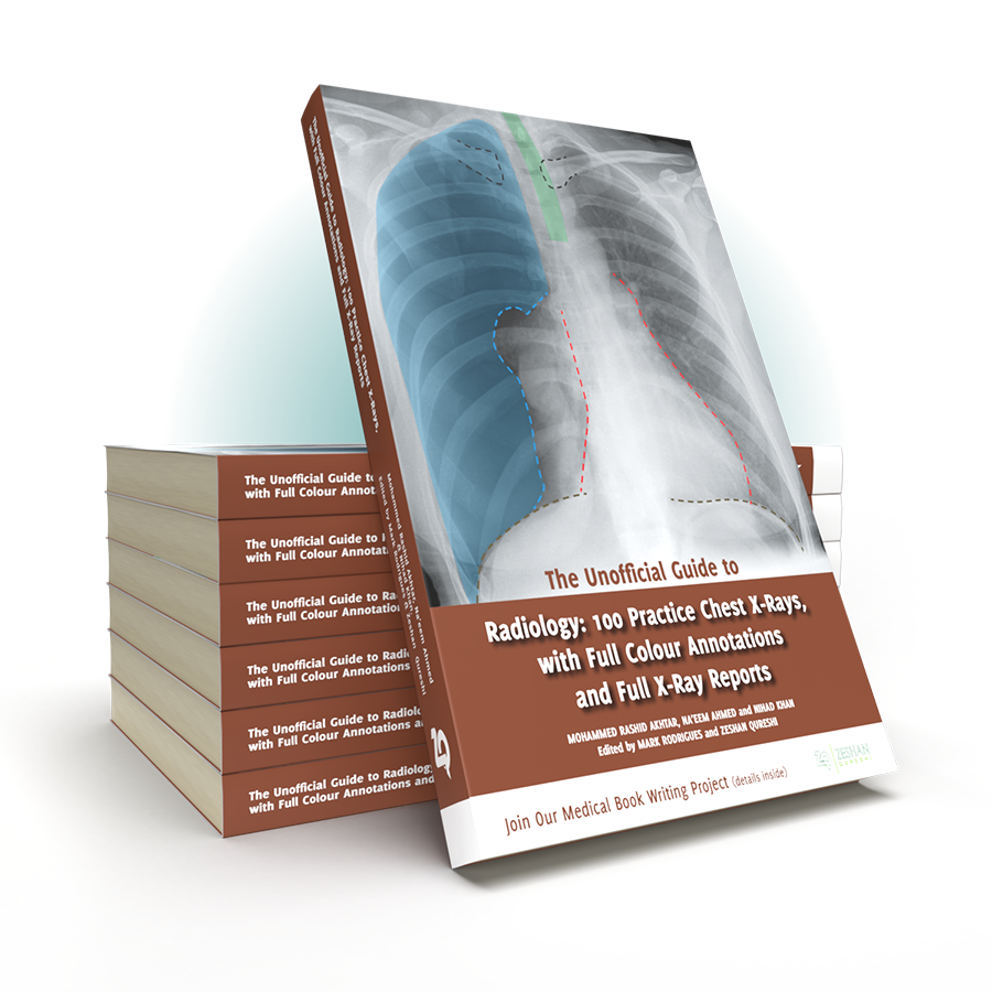
Following on from the multi-award winning Unofficial Guide to Radiology, the Unofficial Guide team return with a practical work book on chest X-rays interpretation, giving you all the practice you need to master this vital skill.
Despite its universal importance, X-ray interpretation is often an overlooked subject in the medical school curriculum, making it difficult and daunting for many medical students and junior doctors.
The Unofficial Guide to Radiology: 100 Practice Chest X-Rays, with Full Colour Annotations and Full X-Ray Reports aims to help address this. This follow-up textbook, specifically designed for medical students, radiographers, physician’s associates, and junior doctors, builds upon these foundations, providing readers with the opportunity to practise and consolidate their chest X-ray assessment and presenting skills.
Key Features
Written in line with the other books in the series, stations contain:

Zeshan Qureshi
Chief Editor
Paediatrics trainee at Great Ormond Street, and the Institute of Child Health
With this textbook, we hope you will become more confident and competent interpreting chest X-rays, both in exam situations and in clinical practice.
We want you to get involved! This textbook has been a collaboration with junior doctors and students just like you. You have the power to contribute something really valuable to medical education; we welcome your suggestions and would love for you to get in touch.
A good starting point is “The Unofficial Guide to Medicine” facebook page, an active forum for medical education. Please get in touch and be part of the medical education project.
100 Chest X-rays: Reported and Annotated JUST LIKE THIS
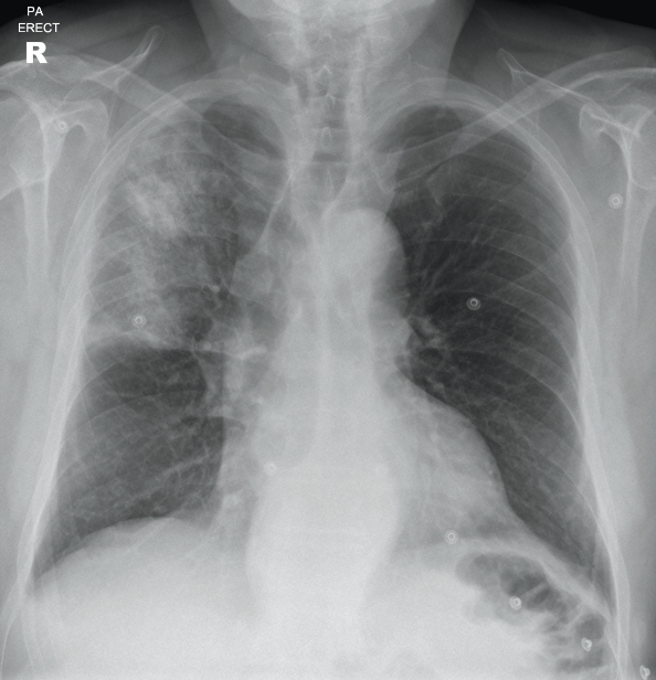
A 70 year old male who lives in a residential home presents to ED with increasing confusion. He has a productive cough and a fever. He has a past medical history of hypertension, angina and mild cognitive impairment. He has a 25 pack year smoking history. On examination, he has saturations of 89% in air, and is febrile with a temperature of 38.8°C. There is dullness to percussion and coarse crackles in the right upper zone. A chest X-ray is requested to assess for possible pneumonia or collapse.
Can you report the X-ray and put in place a management plan?
Turning over the page reveals a full on-image colour annotated X-ray, along with the report, detailing everything you should be expected to see on the X-ray.
Full Report Covering Every Detail Of The X-Ray You Need To Know
Patient ID: Anonymous
Projection: PA
Penetration: Adequate – vertebral bodies just visible behind heart
Inspiration: Adequate – 8 anterior ribs visible
Rotation: Not rotated
AIRWAY
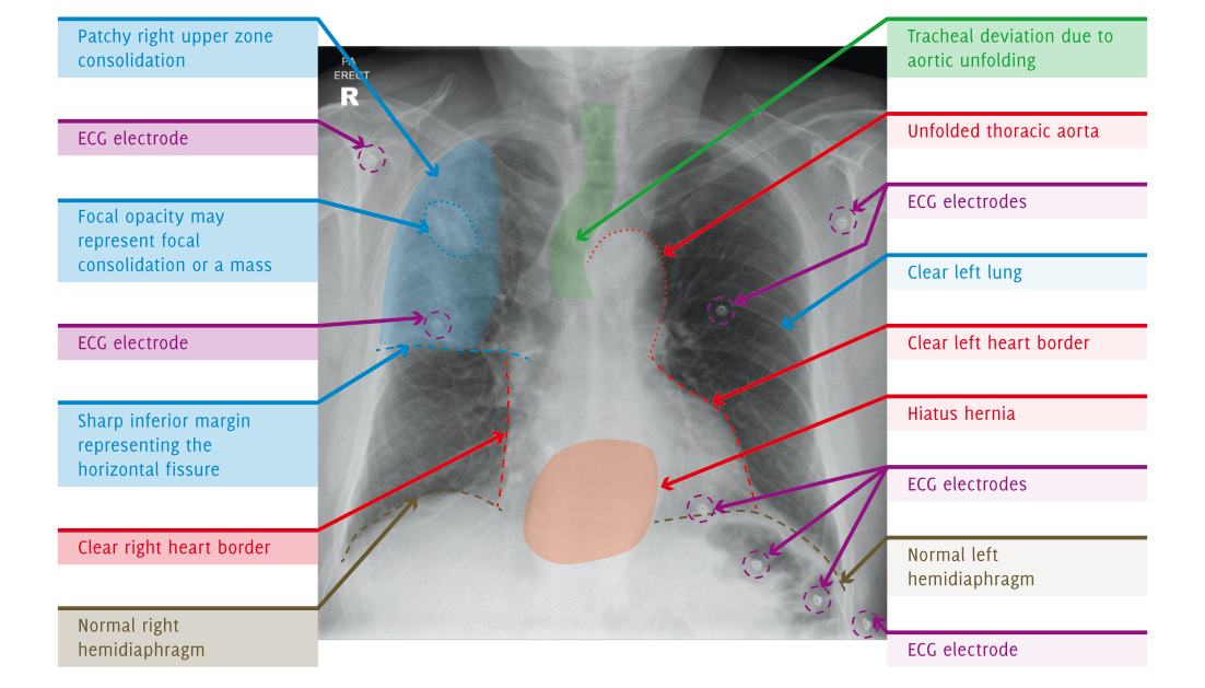
The upper trachea is central. The lower trachea is displaced to the right by the aortic arch.
BREATHING
There is heterogeneous air space opacification in the right upper zone. This has a relatively well defined inferior margin, which is likely to represent the horizontal fissure. There is a focal area of increased opacification in the right upper zone, which may represent focal consolidation or an underlying mass. The remainder of the lungs are clear. The lungs are not hyperinflated.
The pleural spaces are clear.
Normal pulmonary vascularity.
CIRCULATION
The heart is not enlarged.
The heart borders are clear.
There is unfolding of the thoracic aorta, which displaces the lower trachea to the right.
The mediastinum is central, not widened, with clear borders. There is a welldefined density projected over the lower mediastinum, which is in keeping with a hiatus hernia.
Normal size, shape, and position of both hila.
DIAPHRAGM + DELICATES
Normal appearance and position of hemidiaphragms.
No pneumoperitoneum.
The imaged skeleton is intact with no fractures or destructive bony lesions visible.
The visible soft tissues are unremarkable.
EXTRAS + REVIEW AREAS
ECG electrodes in situ.
No vascular lines, tubes or surgical clips.
Lung Apices: Heterogeneous right apical consolidation. Normal left apex
Hila: Normal
Behind Heart: There is a retrocardiac density, which represents a hiatus hernia
Costophrenic Angles: Normal
Below the Diaphragm: Normal
Fully Annotated X-Ray, With On Image, Colour Labeling For Unbeatable Clarity
SUMMARY, INVESTIGATIONS & MANAGEMENT
This X-ray demonstrates heterogeneous right upper zone consolidation in keeping with pneumonia. The consolidation has a relatively abrupt inferior margin in keeping with the horizontal fissure, indicating this is right upper lobe pneumonia. A focal opacity in this region may represent focal consolidation or a mass. Incidentally, there is also a hiatus hernia.
Initial blood tests may include FBC, U/Es, blood cultures, and CRP. A sputum culture may also be taken. The patient should be treated with appropriate antibiotics for community-acquired pneumonia, and a follow-up chest X-ray performed in 4-6 weeks to ensure resolution. The antibiotics may be oral or intravenous depending on the severity of pneumonia (CURB-65).
If the focal opacity in the right upper zone does not resolve then a CT of the chest and abdomen with IV contrast would be appropriate to assess for a lung tumour. It would also be useful to review previous imaging and case notes to see if there was an abnormality at this site before.
Contact Us
Are you a medical trainee, a student, a doctor or medical professional?
We’d love to hear from you for feedback or if you would like to contribute towards the Unoffical Guide to Medicine series.


