Following on from the multi-award winning Unofficial Guide to Radiology, the Unofficial Guide team return with a practical work book on orthopaedic X-rays interpretation, giving you all the practice you need to master this vital skill.
Despite its universal importance, X-ray interpretation is often an overlooked subject in the medical school curriculum, making it difficult and daunting for many medical students and junior doctors.
The Unofficial Guide to Radiology: 100 Practice Orthopaedic X-Rays, with Full Colour Annotations and Full X-Ray Reports aims to help address this. This follow-up textbook, specifically designed for medical students, radiographers, physician’s associates, and junior doctors, builds upon these foundations, providing readers with the opportunity to practise and consolidate their orthopaedic X-ray assessment and presenting skills.
Key Features
100 large, high quality orthopaedic X-rays to assess.
Cases presented in the context of a clinical scenario and covering a wide range of common and important findings (in line with the Royal College of Radiologists’ Undergraduate Radiology Curriculum)
Detailed on-image colour annotations to highlight key findings
Comprehensive systematic X-ray reports
Relevant further investigations and management are discussed for each case
The Unofficial Guide to Radiology: 100 Practice Orthopaedic X-Rays is an invaluable resource for anyone hoping to gain a practical understanding and a wealth of experience of orthopaedic X-rays and their interpretation in a concise, student-focused manner.

Christopher Gee
MBChB MSc FRCSEd(Tr&Orth) MFSTEd
Co-Author
With this textbook, we hope you will become more confident and competent interpreting orthopaedic X-rays, both in exam situations and in clinical practice. We want you to get involved! This textbook has been a collaboration with junior doctors and students just like you. You have the power to contribute something really valuable to medical education; we welcome your suggestions and would love for you to get in touch.
A good starting point is “The Unofficial Guide to Medicine” facebook page, an active forum for medical education
Please get in touch and be part of the medical education project.
100 Orthopaedic X-rays: Reported and Annotated JUST LIKE THIS
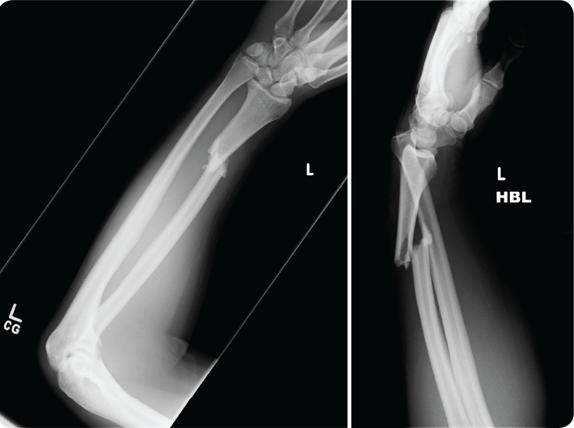
A 20-year-old student presents to the ED, having fallen whilst ice skating. She reports landing on her left forearm. There is no significant past medical history. On examination, there is an obvious deformity with swelling and bruising of the left forearm. The patient is unable to pronate due to pain. Distal pulses are present and sensory and motor function is preserved. The injury is closed. AP and lateral X-rays of the left forearm inclusive of the elbow and wrist are requested to assess for a fracture.

Can you report the X-ray and put in place a management plan?
Turning over the page reveals a full on-image colour annotated X-ray, along with the report, detailing everything you should be expected to see on the X-ray.
Full Report Covering Every Detail Of The X-Ray You Need To Know
Patient ID: Anonymous
Area: Left forearm
Projection: AP and lateral
Technical Adequacy:
- Adequate coverage on the AP view
- Inadequate coverage on the lateral view as it does not include the elbow joint
- Adequate exposure
- The patient is not rotated
FRACTURE DETAILS
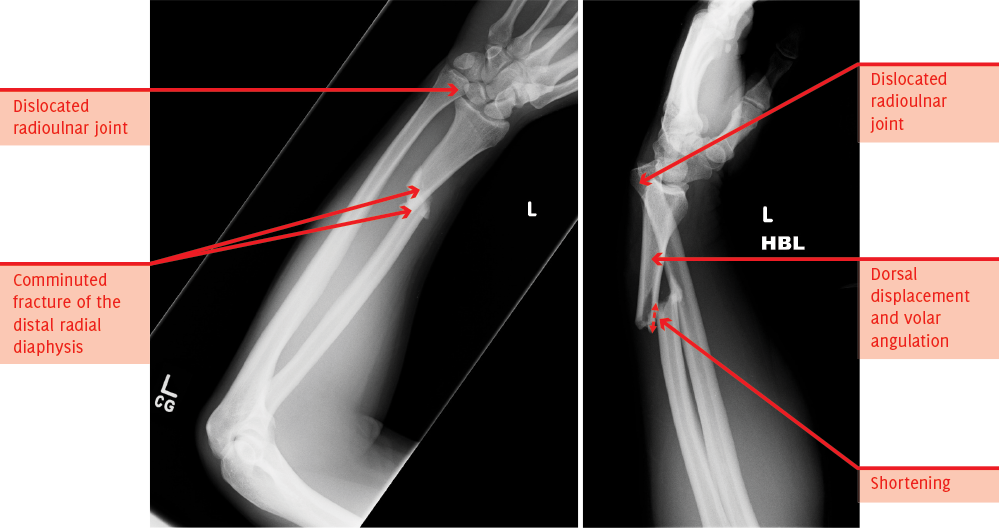
There is a diaphyseal fracture of the distal third of the radius.
The fracture is oblique, comminuted and extra-articular.
There is dorsal (posterior) and medial (towards the ulnar) displacement of the distal fracture fragment.
There is volar angulation of approximately 30 degrees.
There is no rotation.
There is shortening.
JOINTS
There is a dislocation of the distal radioulnar joint.
The elbow joint appears congruent, although this has not been fully assessed.
There are no loose bodies.
There is no effusion or lipohaemarthrosis visible.
There are no arthritic changes.
SOFT TISSUES
There is no soft tissue swelling.
There is no surgical emphysema.
BACKGROUND BONE
The background bone is normal.
BONE LESIONS
There is no bone lesion present.
Fully Annotated X-Ray, With On Image, Colour Labeling For Unbeatable Clarity
SUMMARY, INVESTIGATIONS & MANAGEMENT
These X-rays demonstrate a displaced fracture of the distal third of the radial diaphysis. There is an associated dislocation of the radioulnar joint, consistent with a Galeazzi fracture-dislocation.
A lateral X-ray of the elbow is required.
Appropriate analgesia should be provided.
The patient should undergo attempted reduction in ED under sedation with orthopaedics present. A moulded back slab should be applied and an X-ray taken to check its position. Galeazzi fracture-dislocations require surgery and therefore need early referral to orthopaedics.
Contact Us
Are you a medical trainee, a student, a doctor or medical professional?
We’d love to hear from you for feedback or if you would like to contribute towards the Unoffical Guide to Medicine series.


