Do you have OSCE examinations coming up?
Would you like reinforcement of key clinical signs/tests you are expected to know, with high quality images including real patients with real signs? And do you want to know exactly how to present your findings back to the examiner, and answer the questions they fire back at you?
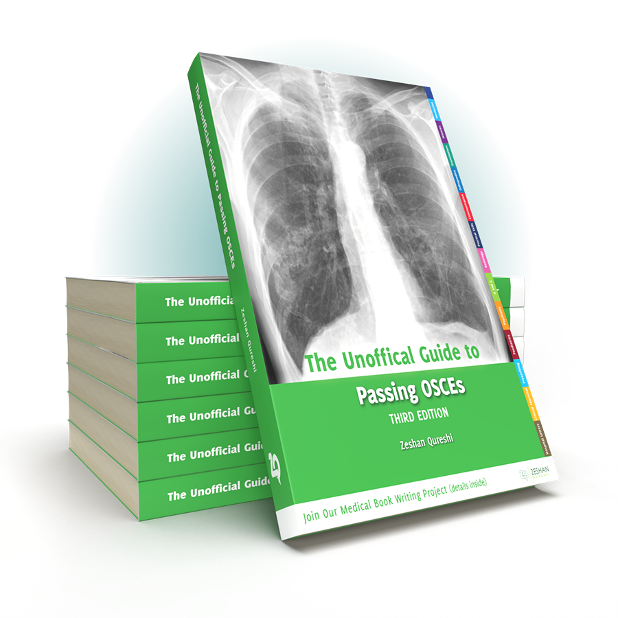
As the best-selling OSCE book on Amazon.co.uk since it’s launch in February 2012, and with copies sold in over 20 countries, The Unofficial Guide to Passing OCSEs is undoubtedly a resource students rely on time and time again to succeed as they progress in their medical career.
In the increasingly competitive world of medical training, the pressure is on to successfully pass one of the most stressful exams you will take at medical school: the OCSE. With its test focused format, The Unofficial Guide to Passing OCSEs features everything necessary to successfully pass this exam. The text captures the insights of medical students and recent graduates in the language that they use to make complex material more easily digestible, with the reassurance of content reviewed by senior clinicians.
Key Features
Unlike other OSCE books, everything in one place. Over 100 OSCE scenarios: histories, examination, practical/communication skills, PLUS specialities including orthopaedics, paediatrics, psychiatry, radiology, prescribing, O&G, opthalmology, ENT
Chapters written and conceptualised by actual students and junior doctors who have recently completed the exam process, in conjunction with over 35 senior clinicians in each specific speciality
Over 300 full colour clinical photos, including real patients with real signs
Model answers to key OSCE questions
Present your findings: how to confidently, concisely and accurately tell the examiner your findings for every station, something feared, yet not covered by other OSCE books, including for ECGs, X-Rays, fundoscopy, and auroscopy
This product stands for quality
All in one place: Step by step guides, over 100 OSCE scenarios: histories, examination, practical/communication skills, PLUS specialities including orthopaedics, paediatrics, psychiatry, radiology, prescribing, O&G, opthalmology, ENT.
The intention of the book is to present key medical knowledge in a manner that is both fun and easy to remember. Over 300 full colour clinical photos, including real patients with real signs. Model answers to key OSCE questions. How to ‘present your findings’ for every station, including ECGs, X-Rays, fundoscopy, and auroscopy. All in the simple language that fresh graduates used to successfully pass exams.

Zeshan Qureshi
Chief Editor
Paediatrics trainee at Great Ormond Street, and the Institute of Child Health
We appreciate that OSCEs are often the most stressful exams you will take at medical school. This books aims to empower your examination preparation and help you on your way to excelling in the OSCE. We believe that fresh graduates have a unique perspective on what works for students. We have captured the insight of medical students and recent graduates in the language that they used to make complex material more easily digestible as students, with the reassurance of content review by senior clinicians.
Every medical student has the potential to contribute to the education of others by innovative ways of thinking and learning. This book is an open collaboration with you: the readers of the second edition have become the writers of the third edition, so please get in touch, you have the power to contribute something valuable to medicine.
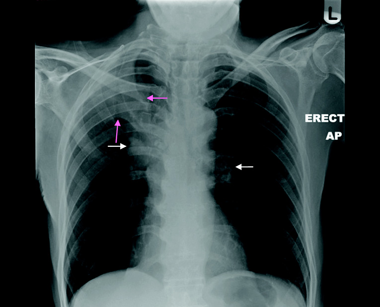
This is an AP chest radiograph. There are no identifying markings. I would like to ensure that it is the correct patient and to check the date that it was taken.
It is a technically adequate film; it is not rotated, has adequate penetration, and good inspiratory effort. There are no important areas cut off at the edges of the film. The most striking abnormality is a well-demarcated area of increased opacification in the right upper zone (pink arrows). The right hilum is increased in size, with a round opacity, and higher than the left (white arrows), suggesting loss of volume. Reviewing the rest of the film, the trachea is not deviated. Comparing the left and right lungs, I can see no other obvious abnormalities in the lung fields. The heart is not enlarged (noting that this is an AP film), heart borders are clear, as are the cardiophrenic angles. There is no abnormality visible behind the heart. The diaphragms look normal, the costophrenic angles are clear, and there is no air under the diaphragm. There is no evidence of fractures. In summary, this chest radiograph shows right upper lobe collapse with associated right hilar enlargement. The most likely diagnosis is neoplasia, although hilar lymphadenopathy may also be due to infection of sarcoidosis.
“This is Mrs Smith, who has presented with shortness of breath and a heart murmur. She is comfortable at rest, on 2 litres of oxygen. There are no stigmata of infective endocarditis. Her pulse is regular at a rate of 60BPM. It is slow rising and normal volume. Pulse pressure is normal. On auscultation, she has an ejection systolic murmur in the aortic area, radiating to both her carotids. S1 is normal with a quiet S2. There are no additional heart sounds. There is no evidence of heart failure.
This is consistent with a diagnosis of aortic stenosis. I would like to confirm this with an echocardiogram, perform a chest X-ray, and an ECG.”
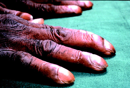
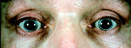
Disease illustrated as you would see it in real life, through pictures of real patients with real signs
Clubbed Fingers
Corneal Arcus
Normal Examination Demonstrated in Pictures
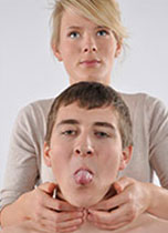
Palpatating for thyroglossal cyst
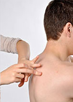
Percussion
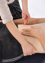
Fluid Thrill
Top tips from both experts and junior doctors
“When introduced to a patient with a murmur, most students will only listen to the murmur on one occasion. I found that, providing the patient was happy, going back and listening to the murmur a second or third time really helped consolidate my pattern recognition skills”
Zeshan Qureshi,
Paediatric Trainee, London
“It is actually much more reliable to look for fasciculations when the tongue is resting in the mouth, rather than when it is protruded.”
Richard Knight,
Professor of Clinical Neurology, University of Edinburgh
Answers to OSCE questions you might be asked
How would you differentiate an upper motor neuron VII lesion from a lower motor neuron VII lesion?
The muscles of the forehead have bilateral cortical representation. Therefore, in an upper motor neurone lesion, e.g. a cerebral infarct, there is sparing of the forehead. The patient would still be able to raise their eyebrows equally on both sides.
How would you confirm the diagnosis of Acromegaly?
Measure growth hormone levels during an oral glucose tolerance test. In acromegaly, the growth hormone level is not suppressed after consuming the carbohydrate load. There may even be a paradoxical rise in growth hormone levels.
What are the most alarming features of depression one should look out for?
Psychotic features e.g. nihilistic/persecutory delusions, auditory hallucinations. Self-neglect e.g. reduced appetite may result in significant weight loss. Potential for harm to self or others.
Contact Us
Are you a medical trainee, a student, a doctor or medical professional?
We’d love to hear from you for feedback or if you would like to contribute towards the Unoffical Guide to Medicine series.
